
Dental X-rays
X-rays can provide information about teeth and the surrounding structures. They are recommended when the additional information is needed for diagnosis or treatment planning.
We offer three types of dental X-rays: small intraoral X-rays, panoramic X-rays and CBCT scans. Each provides a different view, and your dentist recommends the imaging needed for your diagnosis or treatment plan.
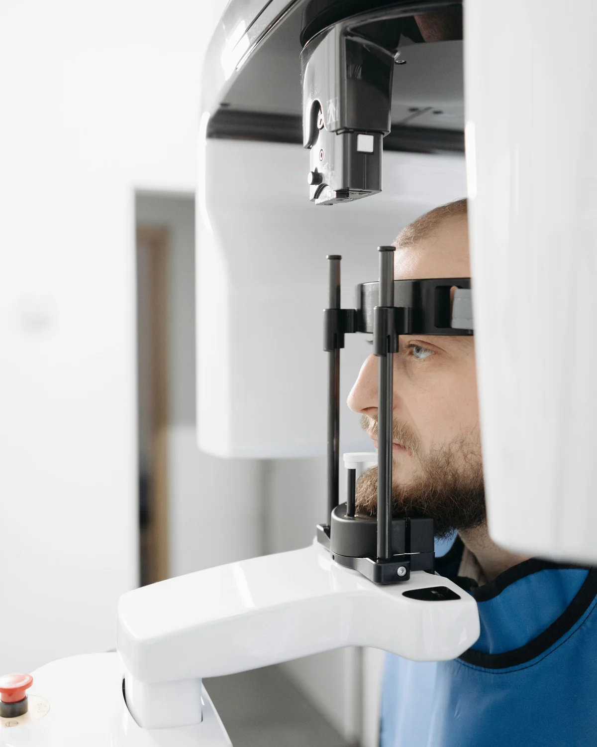
Three types of dental X-rays
Small intraoral X-rays
Detailed two-dimensional images of individual teeth or a small area, taken with a sensor inside the mouth. They help assess decay between teeth, roots, surrounding bone and changes around root tips, and support root canal treatment.
Panoramic X-rays
A broad two-dimensional view of the teeth and jaws in one image. Useful for assessing wisdom teeth, unerupted teeth and the overall arrangement of teeth and jaw structures when a wider view is needed.
CBCT scans
Cone beam computed tomography creates three-dimensional views of teeth, bone and surrounding structures. It can help plan dental implants, assess the relationship of teeth to nerves or sinuses, and investigate selected complex problems when two-dimensional images do not provide enough detail.
Before you begin
Common questions to help you feel informed and ready for your consultation.
Why is an X-ray needed if my teeth look healthy?
An image can show areas that cannot be seen directly, such as between teeth and around roots. It is recommended when the information would help diagnosis or planning.
Should I mention a possible pregnancy?
Yes. Tell the team if you are pregnant or might be pregnant. Your dentist will assess whether imaging is necessary and discuss the decision with you.
Can you use images taken elsewhere?
Tell us about recent dental images and, where possible, arrange for copies to be shared. Your dentist will decide whether they provide the information needed.